Interdisciplinary Research Laboratory (IFL): Researching medicine and technology together
NEWS,
Innovation,
Health,
Research
|
New solutions at the interface between medical technology and machine-assisted processes in medicine: this is the goal of the Interdisciplinary Research Laboratory (IFL), which was set up seven years ago at the Klinikum rechts der Isar, by Prof. Nassir Navab. Currently, the lab is home to over ten research projects running in parallel, where robotics and artificial intelligence play an important role.
The lab’s connection to the Klinikum rechts der Isar was a deliberate choice: Prof. Nassir Navab's high-tech projects at the IFL are collaborative and require the help of medical staff. The interdisciplinary transfer of technical developments into clinical routine takes place step by step, involving constant feedback from the end user and clinic’s medical staff.
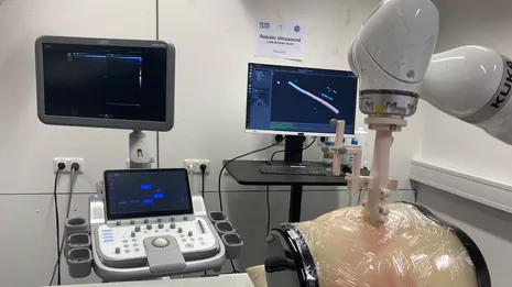
One prime example of interdisciplinary research between computer science and the Klinikum rechts der Isar is in robotic ultrasound: Prof. Navab, head of the Chair of Computer Science Applications in Medicine and Augmented Reality at the Technical University of Munich (TUM), is working on automating ultrasound examinations in the future, without the presence of medical professionals. Such a development would reduce the workload for doctors and save time and medical resources. In practice, 3D images can already show the narrowing of blood vessels in the abdomen or arteries in the forearm. Learn more about these developments here.
Robotics projects: From ultrasound imaging to Loop-X
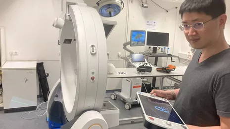
At the IFL, about 30 researchers are focusing on several automation and robotics projects, including the automatic creation of reports based on X-ray images, and the use of the mobile imaging robot "Loop-X" (from Brainlab) for flexible computed tomography (CT), a significant component in robot-assisted ultrasound imaging.
Scientist Dr. Zhonglian Jiang is responsible for robotics projects at the IFL. The following projects are of particular interest here:
"Robotic Ultrasound for Spinal Injections": a project developing a robotic system that supports doctors in safely injecting into the spine. Independently from human experts, the system finds the region in which the injection can be safely made, and shows ultrasound images of the region.
"Intelligent Robotic Sonographer for Autonomous Scanning": Researchers are developing a way to automate real-time diagnosis of vascular diseases using ultrasound. To do this, it is important to place the ultrasound directly on the skin with the right amount of force, precisely targeting the arteries. This can be better assured with automated processes – another strength of the developed method is the strong reproducibility of its results.
"Optimal path planning for autonomous robotic ultrasound scanning (reinforcement learning)": This project aims to make more efficient and effective the visualization of structures by the ultrasound head. For example, various rotation and translations may be required to eliminate noise from a produced image, for example from shadows caused by the ribs. This technology can improve the overall image quality by finding optimal locations and paths for the probe.
Around 30 researchers are employed in Prof Nassir Navab's Interdisciplinary Research Laboratory IFL.
To learn more about what is happening at Prof. Navab's lab, take a look at these videos showcasing the lab's current robots and possible applications in clinical work 🌟